Saw this movie today on the BBC news website: The World's Smallest Movie. It's made using the scanning tunnelling microscope - a close relative ('the parent') of the atomic force microscope. Each 'ball' is a carbon atom that is moved around on a copper surface. The BBC website also uses 'the diameter of a human hair', again, as a length comparison: 1000 frames (images) would span a hair's width. ~James
Using the literal/self model, which has its restrictions, proposed in a previous blog entry to aid/classify the interpretation process, I thought about some possible artworks that could be produced - mainly at the literal ('science-end') of the spectrum of interpretations. I might have a go at some of these myself using the micrographs directly or by manipulating photographs. The ideas are listed below. They may also serve to give further inspiration to others; I haven't really thought about the 'self-type' interpretations, although some of these ideas may provide pointers to this realm.
~James 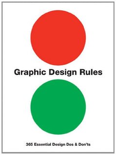 I was in Waterstones bookshop today and a book caught my eye: 'Graphic design rules. 365 Essential design do's and don'ts' by P. Dawson et al. Rule number 178 stood out to me: 'Thou shalt remember that people like bright things - brown is not exciting.' It made me laugh because the default palette for AFM images is brown. All the hair images I gave out last week to the TAG members were brown.
Whilst writing this, my wife reminded me of a lady who rang up to complain about travelling on a brown coach. She returned on a more colourful coach. ~James The schematic below provides a summary, with an example, of the kind of image interpretation I had in mind, although I'm sure there are many other ways of looking at it. ~James 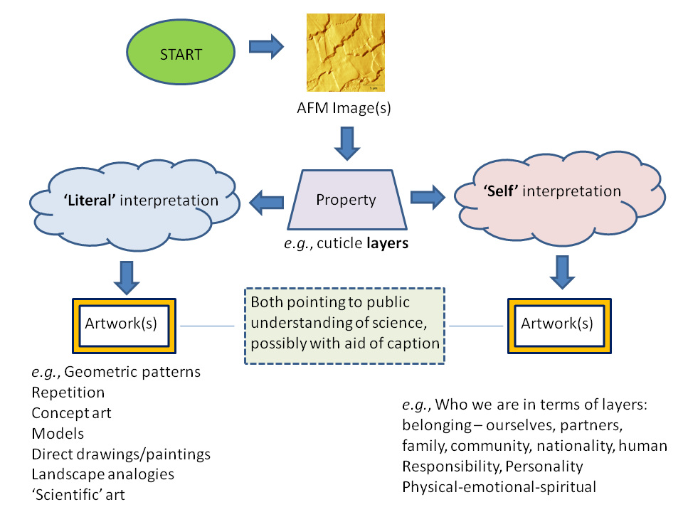 Saw this picture on facebook and it reminded me of some 'flythrough' movies of AFM images that University of Bristol did in the mid-1990s. This type of thing is possible since AFM images contain 3D info: length, width and height. Maybe there's some free software 'out there' for doing this now... will investigate. ~James 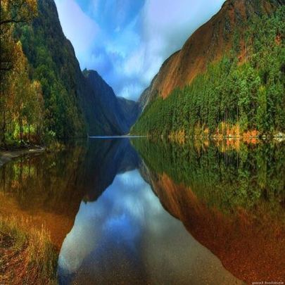 Mountain Lake, Glendalough, Ireland
Ilaria Vanzulli, a PhD student in the School of Pharmacy of Biomedical Sciences at the University of Portsmouth, produced this comparative image from some of her cell samples. I saw the image on facebook and she agreed that I could share it here. It seemed quite fitting. ~James 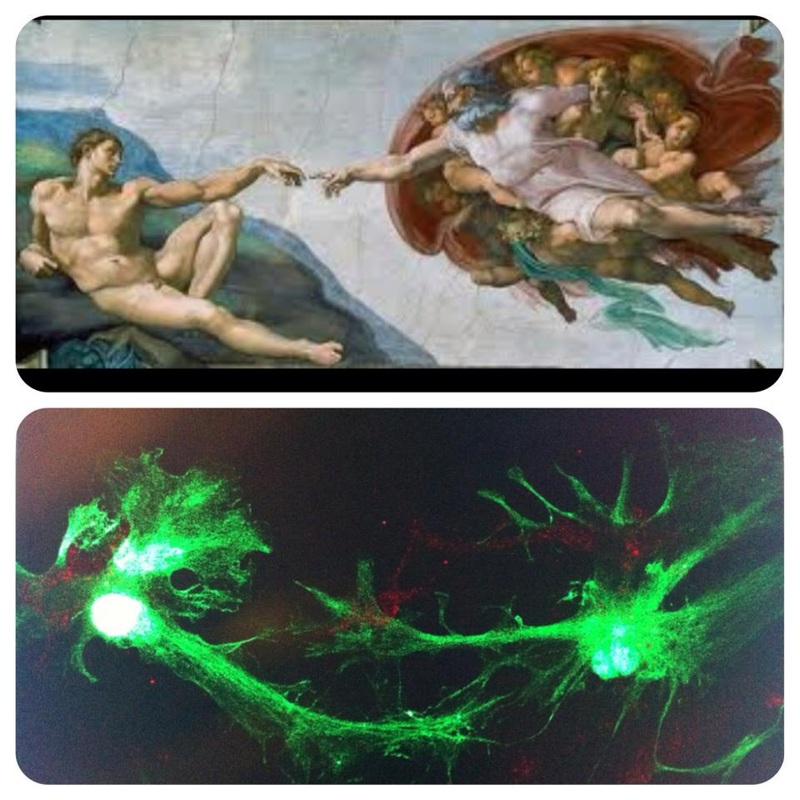 (C) Ilaria Vanzulli and Michelangelo
There are many examples of combining art and science/microscope images, some of which I'll post here in due course. The petri dish paintings by Klari Reis and their motivation, however, seem to capture a 'response to science' that I had in mind for the project. In other words, the art is not necessarily trying to reproduce the science (micrographs) using different media, which is interesting in it's own right, but add 'something': in this case, Reis's diagnosis with Crohn's Disease. Having said that, I think 3D accurate models would also be welcome: the other end of the spectrum. No doubt the responses will be varied. ~James 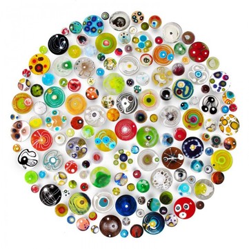 Looking through the University of Swansea 2012 Research as Art competition winners on the Guardian website. Thought the Knitted Protein Model (Josie Parker, College of Medicine) was particularly inspiring. ~James 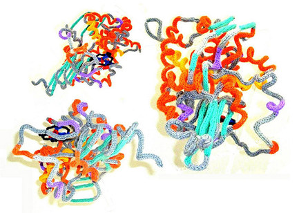 | AuthorsContributions from various team members participating in the project. ArchivesMarch 2014 CategoriesAll |
 RSS Feed